
Ocular Ultrasound
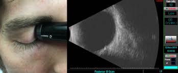
Ocular ultrasound is a technique in which a small ultrasound probe is used to image the eye. Sound waves transmitted from the probe reflect differently through various layers of the eye, thus generating a virtual image. Conditions such as a dense cataract or vitreous hemorrhage can prevent a direct view of the retina. When a view to the inside of the eye is not possible, the ultrasound can produce an image of the entire eye.
How is the test performed?
An ultrasound probe is coated with ultrasound jelly, then gently placed on the outside of your eyelid. It does not need to touch the eye itself. There is no pain in the procedure and no risk of injury. You do not need to be dilated for this procedure, although you may need to be dilated for other parts of the eye exam.
Why do I need this test?
Dr. Blem may recommend this test if his view to the inside of the eye is poor. He may also recommend this test to measure the dimensions of certain intraocular lesions or to assess certain optic nerve related conditions.